Seeing your baby for the first time can be a heart warming moment, and no doubt you don’t want to wait a whole 9 months to be able to do so, which is why many parents start thinking about a 3D ultrasound. However, before you book anything you should know a little more about what a 3D ultrasound is used for, when it is recommended and whether it is safe for you and your baby.
What is the difference between 2D and 3D ultrasounds?
Many people have undergone an ultrasound sometime in their lives, and this is particularly true of women who have passed their 8th week of pregnancy. A 2D ultrasound can be performed in most practitioner’s offices or at the local hospital and provides an exciting first glimpse of your child.
When you are undergoing a 2D ultrasound you will be asked to lie on your back and expose your belly. The practitioner will then rub a gel on your abdomen to help the transmission of sound waves into your body. Once the gel is applied they will use a hand held device called a transducer and rub it against your belly, allowing sound waves from the device to enter your body and bounce off the various structures inside, including your growing child! These sounds are then converted into electrical signals and translated to pictures on a screen.
If the ultrasound is very early on in your pregnancy then an abdominal ultrasound may not be possible. In that case it would be performed by a vaginal probe, rather than hand held transducer.
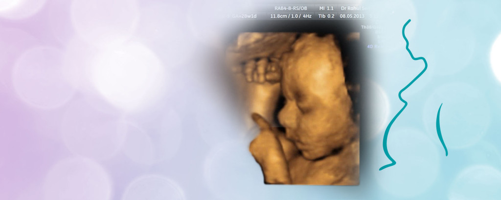
A 3D ultrasound is similar to a 2D ultrasound, except multiple two-dimensional images are taken at different angles, and then pieced together to provide a three-dimensional image of the child.
What are 2D and 3D ultrasounds used for during pregnancy?
2D ultrasounds are a regular part of the prenatal tests performed during pregnancy. They are normally performed once in the first and once in the second trimester. The first trimester ultrasound is used to pick up the heart rate and see the umbilical cord, size and placenta. The second trimester ultrasound is used to observe the placenta and different features of the foetus including the head, face, spine heart and abdomen. The main reason for these tests is to look for physical abnormalities in the development of the child.
3D ultrasounds are only recommended by the doctor if they suspect a foetal abnormality such as a cleft lip and are not a regular part of prenatal exams. Parents on the other hand sometimes wish to perform a 3D ultrasound for the sake of creating a keepsake of their child, a 3D image that they can show to their friends.
Are 3D ultrasounds safe for the mother and child during pregnancy?
There are many ‘prenatal portrait centres’ that offer 3D ultrasound pictures of your child, however, the safety of these sessions is uncertain. It is recommended by doctors that while ultrasound technology for the most part is considered safe, it should only be used by a trained practitioner and only if it is medically necessary, such as to examine foetal abnormalities.
3D ultrasounds use sound waves which can warm up the tissue a little, which can sometimes create pockets of gas. While there are no known risks with ultrasound technology, the long term consequence of this effect are unknown, so it is always best to be safe.
Therefore, experts discourage the idea of using 3D ultrasounds for creating a keepsake or just to have a portrait of the child. Instead they counsel that they be used only for medical purposes, by a trained professional and only if it is recommended by trained medical staff.